
There is greater awareness among people today regarding the early detection of breast cancer. The increased awareness has led to the resurgence of mammography use. That is why there has been an increase in the number of mammography system installations and also in the number of manufacturers.
Mammography is a technically exacting procedure. Even a small change in technique or processing factors could impact the image quality and radiation dose that is delivered. The focus is always on producing mammograms with the lowest possible radiation doses consistent with high specificity and diagnostic sensitivity. That is why a lot of attention goes into the choice of equipment, imaging techniques, patient positioning, and the development of a quality control program.
Methods of imaging
There are two types of image receptors that are being widely used, high-resolution film-screen systems and xeroradiography.
In high-resolution film-screen systems, there is one thin highly-absorbing intensifying screen with a single-emulsion film. However, a dual-screen image with a double-emulsion anti-crossover film has also been introduced recently.
In xeroradiography systems, a more penetrating X-ray beam is used compared to film-screen mammography. It displays more enhanced images with high spatial resolution and imaging a wide range of tissue attenuation. An electrostatic process produces images using charged toner particles that are attracted to various charged areas on a selenium plate. These particles are then transferred to a background of white reflective paper. The toner particles follow distorted trajectories caused by fringe films above the selenium plate, resulting in the edge enhancement effect.
Whether to use a film-screen or a xeroradiography system depends on many factors, including:
X-ray equipment
In film-screen systems, you will need a special thin window x-ray tube that typically incorporates a molybdenum target and filter. It also has a generate to operate at low voltages. In xeroradiography systems, there is usually a small focus tungsten tube with an aluminum filter. It also comes with a generator that works in the 42 to 52 kVp range.
Patient positioning
For film-screen mammography, it is recommended to use mediolateral oblique and craniocaudal views. Xeroradiography uses medio-lateral oblique and craniocaudal views.
Film-screen mammography also used a grid to improve contrast, but a grid is not needed in xeroradiography.
Film-screen mammography also requires firm compression of the breasts, whereas xeroradiography can tolerate fewer firm compressions due to its wide recording latitude.
Diagnostic image quality
With film-screen imaging, you get better images of soft tissue density tumors. On the other hand, xeroradiography is more useful in the visualizations of microcalcifications and masses that have small radiating fibers or well-defined borders.
Convenience and personal preference
Viewing ease
Xeroradiography images are viewed by reflected light under normal illumination conditions. For clinicians, this is more convenient.
You will need low ambient light on a masked view box to view subtle contrast on film-screen images. You may also require bright lighting to view the darkest regions of the film.
It may also become necessary to use a magnifying lens to view fine structures in both techniques.
Reliability
Downtime and reliability may vary between the two techniques. The xeroradiography system is more complex than a film-screen unit and requires regular maintenance.
Backup processors
Most facilities have more than one automatic film processor. For mammography, it is recommended to use a dedicated processor, and so a second unit can be used in emergencies.
With xeroradiography systems, you are less likely to have an alternate processor. If the xeroradiographic processor is out of service, this may affect system downtimes significantly.
Radiation dose
The average radiation dose for non-grid film-screen imaging is two to three times lower than the xeroradiographic system. The increasing use of grids for film-screen imaging has led to an increase in radiation dose. However, there are now faster film-screen systems available in the market that can compensate for the increased doses when grids are used.
Imaging systems are quickly evolving, which means dose comparisons will change with time.
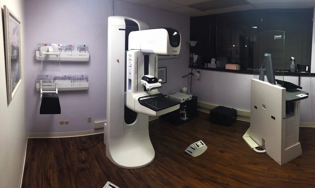
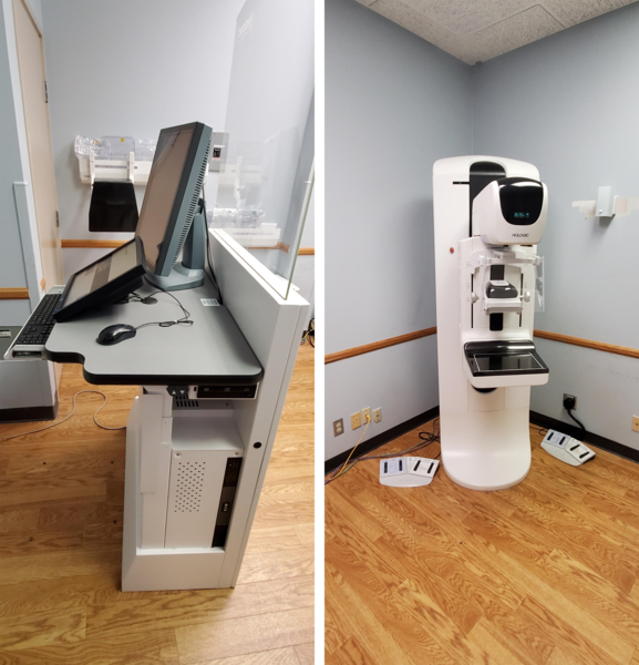
How to prepare for installing a mammography unit?
Proper planning is very important in successfully installing a digital mammography unit. It can help avoid potential issues such as room size, power, HVAC systems, cable drops, and more.
Following these steps can help you in successfully installing a mammography unit:
– Analyze the drawings of your space with your project managers and engineers. Doing so will ensure that you have the right-sized space and also the proper utilities necessary to operate a mammography unit.
– You will also need to have proper power hook-ups, and so consulting an electrician is also a must.
– Ensure that the room you plan to install the mammography unit is cleared before the day of delivery. You should also be aware of any narrow doorways that may hamper the transportation of the unit.
– Check with the vendor regarding the delivery of the mammography unit. You need to ensure that there are no scheduling conflicts.
– To install the mammography unit, you will need to schedule an engineer visit. Typically, an engineer visit for installation happens a couple of days after delivery.
– If you have also purchased a CAD unit with your mammo, connecting it to PACS (Picture Archiving and Communication System) can be a bit complex. Things will get easier if you have the network information prepared in advance for smooth installation.
– You will also need to contact your local radiation authority so that they can send a radiation physicist to inspect your system. Once you receive their approval, your system is ready for use.
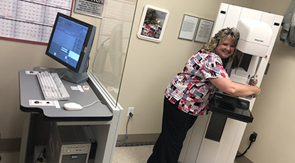
After the engineer has completed the installation, they will generally demonstrate to your technicians how to use the system. However, you may also need to arrange for Application Training that educates your technicians about procedures, quality control protocols, and more. For that, you may need to certified applications training specialist.
Once the engineer gives the demonstration to your technicians, the system is signed for and handed to you. If you have purchased service coverage, it will go into effect from this point onwards. Your unit is ready to scan patients, depending on the requirements of your state for radiation physicist approval.
Different sites may encounter different difficulties in successfully installing a mammography unit. However, proper planning in advance can help you mitigate most issues, resulting in a successful installation.