
New technologies for breast imaging are constantly being developed, and it is important to stay up-to-date. The “Hologic Dimensions: Field Service + BioMed Accelerated Seminar” is a four-day comprehensive training course that covers the fundamental aspects. Each day is divided into two blocks to help participants better understand the concepts.
The training is presented by mammo.com (Powered by UMAC Radiology Sales and Service).
Here is a day-by-day break-down of all the topics covered:
Day 1
First Block – Basic Operations
– Caltools
– System Tools
– Disassembly
– Cover removal
– Detector removal
– Drawer removal
– Computer removal
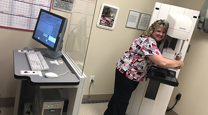
The first block of the first day is dedicated to helping participants understand the basic operations of a mammography unit. Hologic systems have been the leader in breast cancer screening and introduced the first breast tomosynthesis technology, superior to prevailing 2D technology. The mammography systems by Hologic offer exceptionally sharp images and seamless transition between various imaging modes. Hologic systems the first in the world to offer tomosynthesis-guided biopsy.
Participants will learn about the various tools and components used in Hologic’s Mammography Systems. In this course you will also learn how to disassemble the entire system, including cover removal, detector removal, drawer removal, and computer removal.
Second Block – Basic installation
– Mounting
– Cabling
– Power checks
– Alignment checks
– Generator checks
– AEC calibration
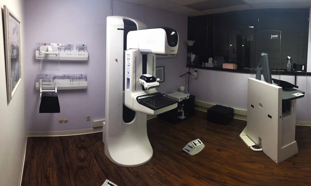
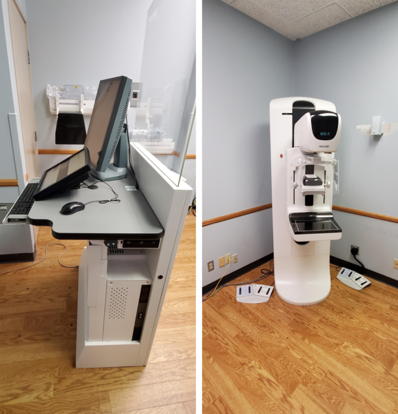
Mammography machines are usually of free-standing design, and so it is important that the system is installed properly and is stable. The control unit needs to be level while allowing enough space for service and maintenance.
The second part of the day will focus on helping participants understand the basic installation procedures for mammography systems. This includes the proper way to mount the system and ensure that all the cables are attached correctly. Participants will also learn to conduct necessary checks, such as power checks, alignment checks, and generator checks to ensure that the unit runs optimally. They will also be taught to check AEC (Automatic Exposure Control) calibration to adjust to breast thickness. Improper AEC calibration can lead to difficulty in creating digital images of consistent optical density.
Day 2
First Block – Tube/Generator Service
– Replacement
– Alignment
– Calibration
– Half-value layer
In mammography, the x-ray tube, the electrical generator, and the overall exposure configuration are particularly important. Unlike traditional radiography, mammography is performed in low kilo-voltage for obtaining adequate tissue contrast. That is why the mammography generator must be accurate and consistent in producing low kilovoltage.
The X-ray tube assembly is similar to that is conventional radiography. However, large amounts of heat are produced, which, if not dissipated, can damage the tube.
In the first block of the second day, participants learn about tube and generator services, including replacing them and aligning them. They also learn about calibration and the half-value layer. The half-value layer is the thickness of a material at which the intensity of radiation entering it is reduced by half.
Second Block – Detector Service
– Replacement
– HVPS replacement
– Upgrading/Downgrading
– AEC refresher
Digital mammograms use solid-state detectors to record the x-ray pattern passing through the breasts instead of film. The detectors convert x-rays passing through them into electrical signals and then send them to a computer.
The second block of the day focuses all on detector services. Participants learn about replacing detectors and HVPS (High Voltage Power Supply) replacements. They will also go through upgrading and downgrading as well as AEC refresher.
Day 3
First Block – C-arm service
– Cage removal
– Compression motor replacement
– Calibration
When installing a mammography unit, it is important that the C-arm translation motion and the rotation of the same be smooth and easily locked in the desired position. The image receptor compartment must be able to secure the image receptor in the right position, and yet, the radiographer must be able to insert and withdraw it easily.
Effective compression is also extremely important in mammography. It causes the breast tissue to be expanded over a larger area, which reduces the thickness. This gives better contrast and, therefore, better imaging.
The third day will start with participants learning about the C-arm service. They will learn to remove the cage as well as replace the compression motor. They will also learn about proper calibration to ensure that the machine runs and operates properly.
Second Block – AWS Computer Replacement
– Computer types
– Important files
– Backup/Restore
For many decades now, researchers have focused on bringing new technologies to the medical imaging field. Researchers have tremendously helped the medical industry by helping doctors spot diseases and identify health issues. Software designers are also playing a key role by creating software that helps doctors make sense of a huge volume of non-invasive medical information.
Radiologists have to look at hundreds of x-rays, MRIs, and other images every day. They are required to identify nuances in hundreds of healthy images, something that could tire even the most disciplined minds.
Companies are therefore working to develop CAD systems to help radiologists improve their efficiency and accuracy in diagnoses.
The second block of this day will focus on the various computer types that are used in different Hologic mammography systems. Since everything is digital, it is imperative that participants understand the various types of computers being used and how to service or replace them. Now, computers do not run without software and files. This section of the training will cover all the important files that are necessary to run the mammography unit.
Sometimes, files may get accidentally deleted or corrupted. Participants will learn the importance of backing up important files and how to restore them if they go missing or get corrupt.
Day 4
Final Day
– Entire Syllabus Recap
– Questions/Discussions
There will be no new topic discussed on the final day. The day will be dedicated to recapping the entire syllabus so that participants can make the best use of the training. Therefor, The day will be reserved for discussions on the topics covered as well as in answering questions from the participants.
If you want to participate in the Hologic Dimensions: Field Service + BioMed Accelerated Seminar, you can call 855-3D-Mammo or email us at hello@mammo.com
Click below to download Syllabus PDF
Mammo_syllabus_v3.3 (1)